
See how River Project Scientists Study Signs of Spring in the Hudson River
Every year, we see the first signs of spring in the life around us — the grass begins to grow, trees start budding and springtime birds return to the city. Under the waves of the Hudson River, similar changes take place in a beautifully important event called a plankton bloom.
As the water begins to warm and nutrients become available, aquatic producers called phytoplankton begin to photosynthesize, exploding in populations as they produce the oxygen and food that organisms higher in the food chain rely on.
After an initial bloom of phytoplankton comes a bloom of zooplankton, animal plankton that rely on phytoplankton (and sometimes other zooplankton) as a food source. This population boom helps kickstart the River’s food chain each season. Because we know the health of plankton populations directly impacts the health of fish, birds and other species higher up on marine food chains, it is important to study this group of organisms and how they are impacted by pollution, climate change and other human activities.
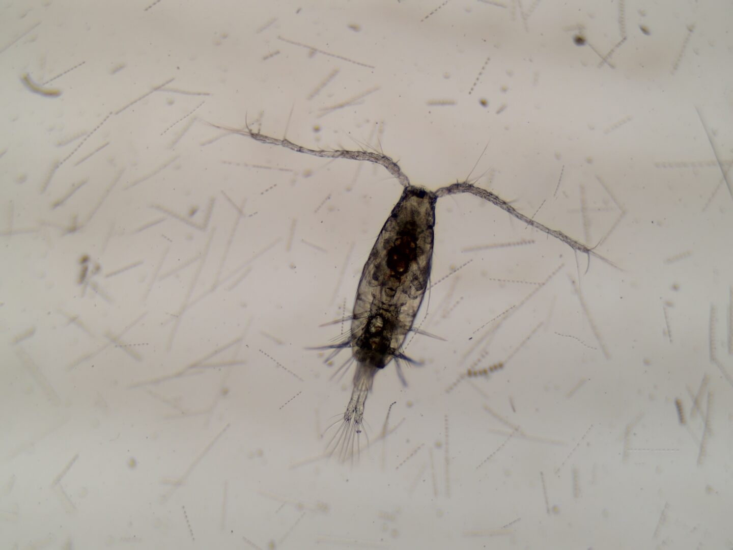
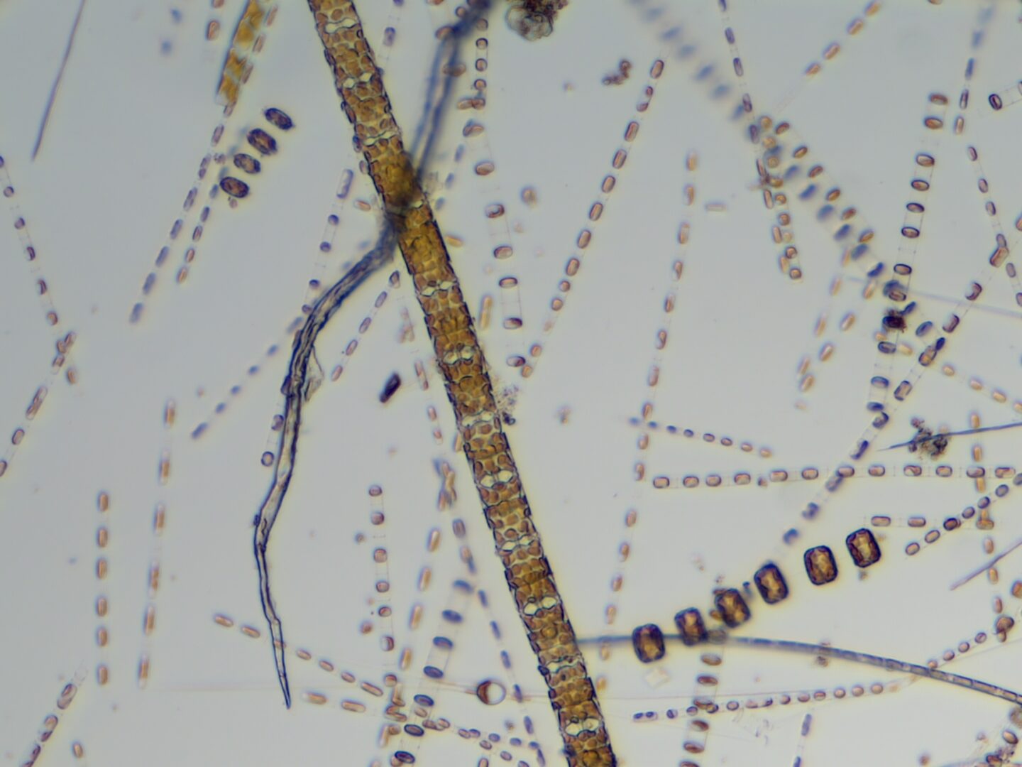
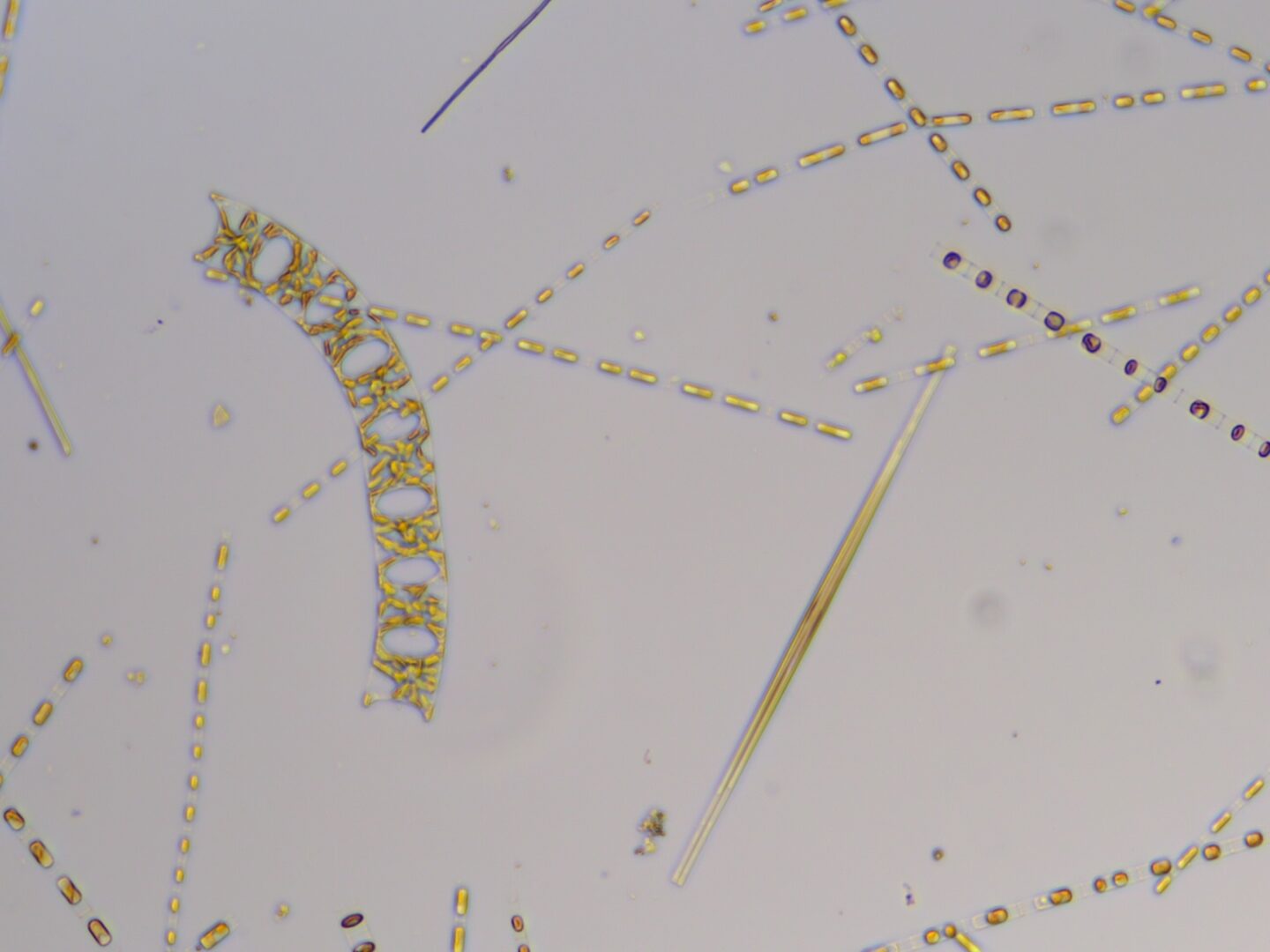
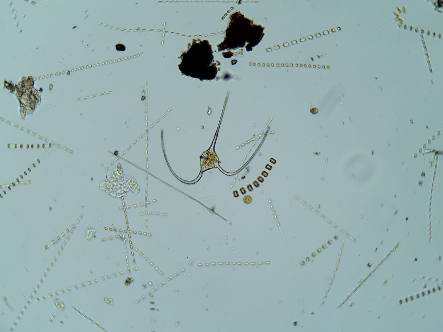
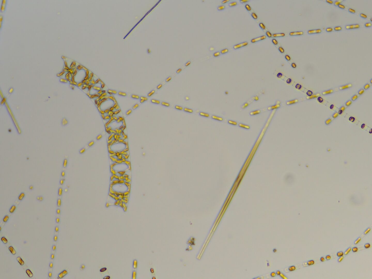
Here, you can see pictures of microscopic plankton captured by our team in our efforts to identify and study these important organisms.
To capture well-defined images with high concentrations of plankton, our River Project team uses special tools and techniques. Today, we’re sharing a look behind-the-scenes (or in this case, the microscope lens) at how we collect population samples, the tools we use to view these tiny organisms and how we go about the sometimes tricky work of species identification.
The plankton shown in these images are tiny. Even though the plankton population in the Hudson River is dense enough to make the water appear green, if we look at a regular water sample under a microscope, the plankton are fairly spread out. This means that it takes a much longer time to find different plankton species to photograph. To get over this hurdle, we use a tool called a plankton net to acquire a concentrated plankton sample.
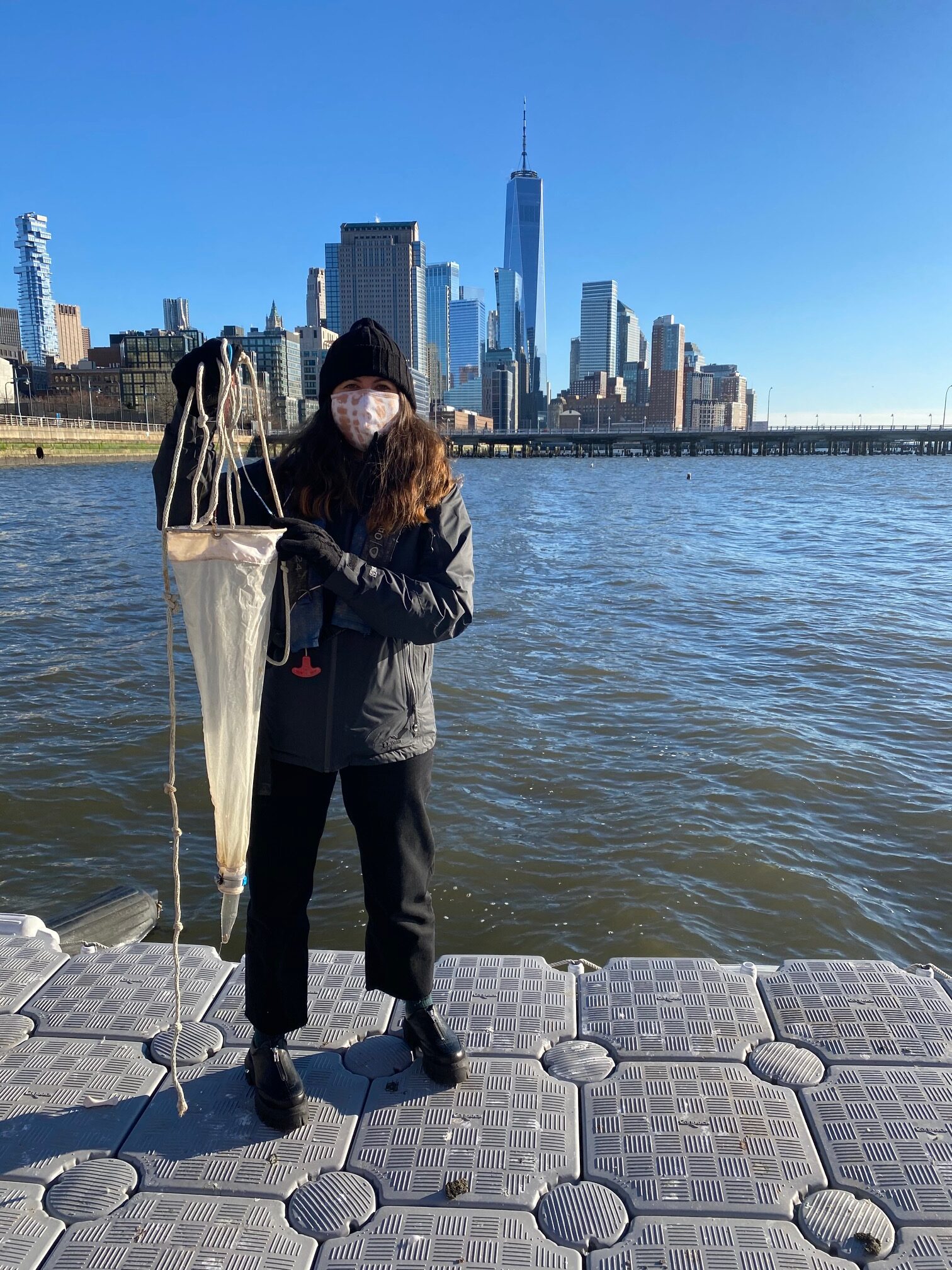
A plankton net is similar to a net that we use to catch fish or butterflies, but the holes in the net are much much smaller. The holes in our plankton net range from 100 micrometers by 100 micrometers to 250 micrometers by 250 micrometers. To put this in perspective, there are 1,000 micrometers in every millimeter, and 10,000 micrometers in every centimeter.
When taking a sample with our plankton net, the plankton that are too large to fit through these holes are caught by the net while the water they float in is able to pass through. As we sweep the net back and forth in the water, we begin to concentrate the plankton in our net, catching them and the water keeps flowing through. Once we bring the net back onto our dock, we collect the captured plankton in a small plastic bottle at the end of the net. This bottle allows the plankton to remain in the water after being collected, before we transfer the sample to one of our glass jars.
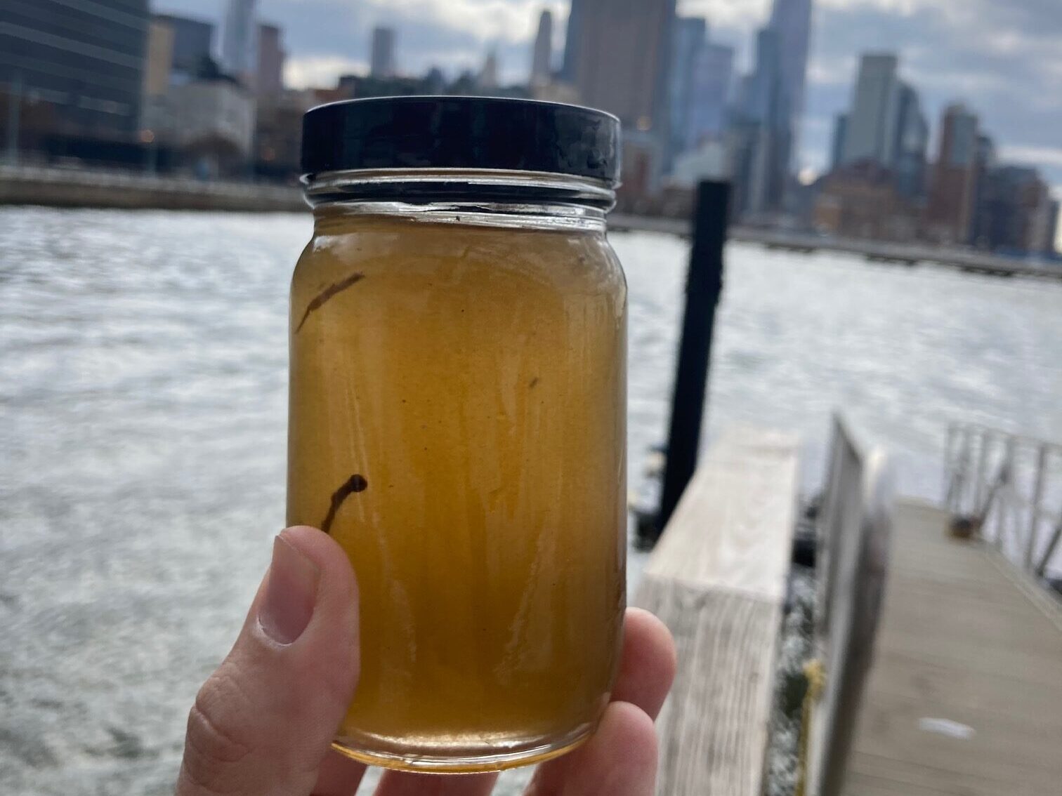
The cloudiness of the water in the same pictures here shows that we have a higher density of plankton per volume of water than we see in the wild. This will help us at the next step in the process — as we look at our samples under the microscope. Concentrated samples deliver more diversity without having to look through as much water, giving us more time to take photographs and identify species!
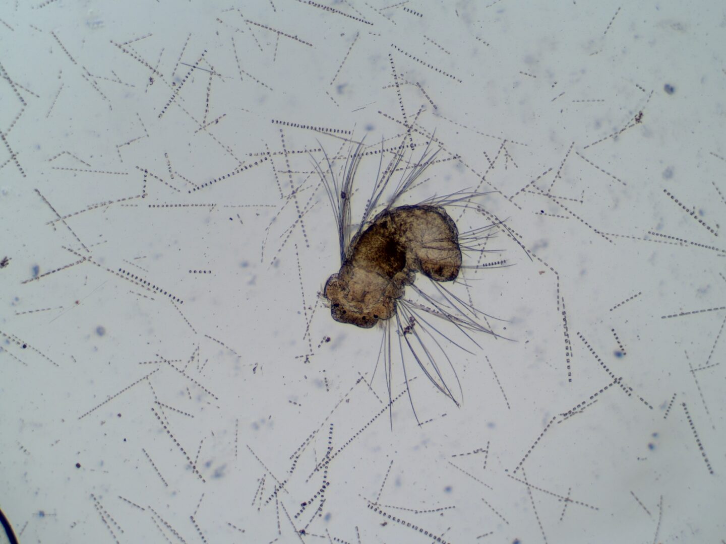
We took all of the plankton images you see here using various cameras mounted onto a compound microscope. A compound microscope is a microscope that makes use of two sets of lenses to achieve a high degree of magnification on microscopic organisms and objects like plankton, allowing us to see features much too small for the unassisted eye to make out. For example, the copepods below might look like a small speck when we look into a plankton sample with our eyes, but under the microscope we can see their legs, antennae, eyespot and other parts of their complex anatomy!
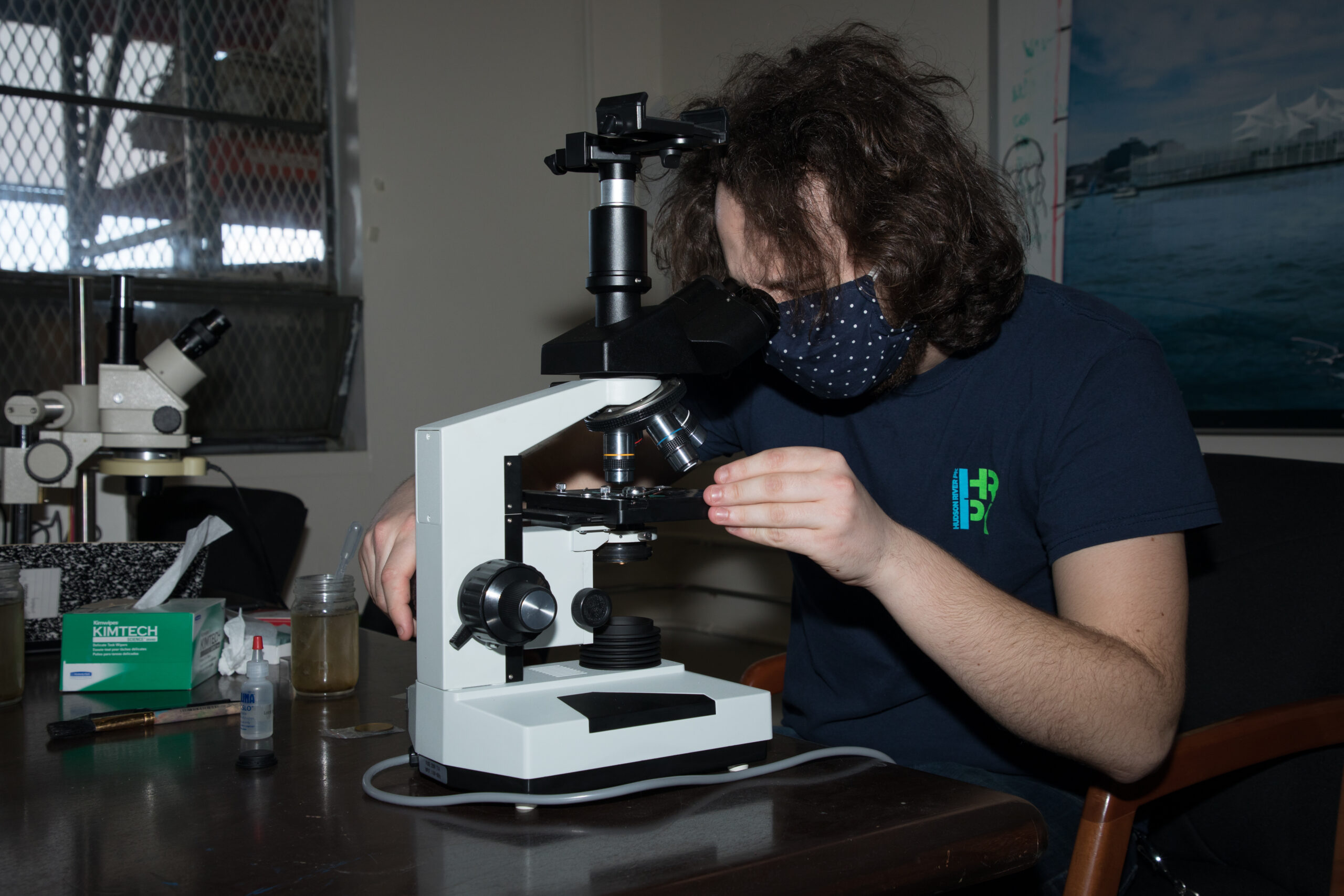
Mounting cameras to our scope enables us to take pictures of exactly what people are seeing when looking through a microscope at a plankton sample. For education purposes, this is incredibly useful as it allows us to all observe the same organism and work together to identify what we are seeing.
So how do we prepare the water we collected with our plankton nets to be viewed and photographed under a microscope? First, we use a pipette to take a drop of water from the sample and place it onto a slide, a small rectangle of glass that we place under the microscope. Depending on how much plankton are in the sample, we may choose to add an extra drop of normal river water to make seeing individual plankton easier. Then we can adjust our lighting, focus, magnification and other microscope settings to move the slide around, see smaller and larger objects, and otherwise get a clearer picture of the organisms and objects under the scope. After that, surveying a slide is similar to birdwatching: you never know exactly what you will find until you go looking!
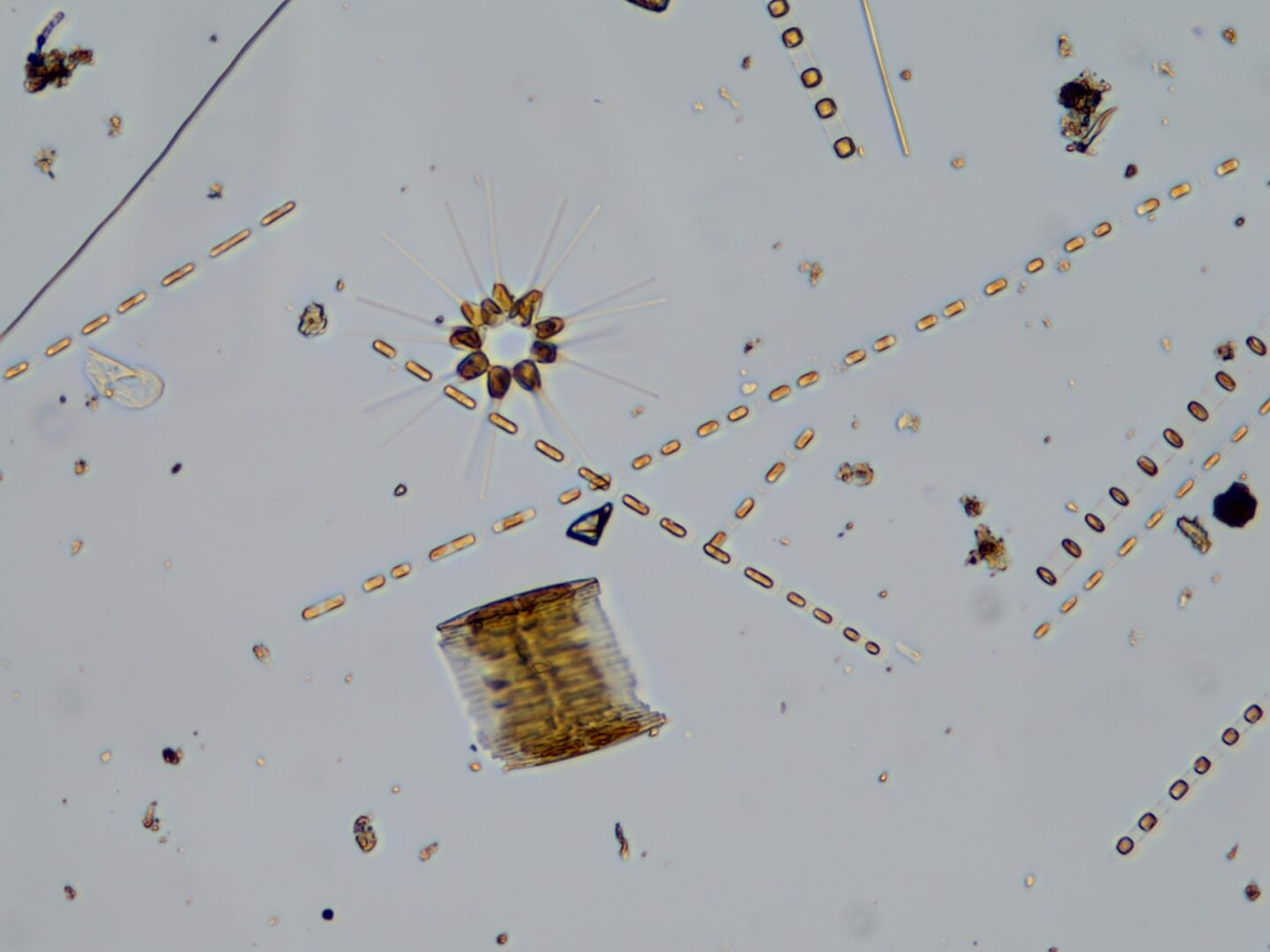
Identification of objects under the microscope can be difficult. Often, we see the organisms from only one field of view, and certain features can be hard or impossible to observe. Generally speaking, the more clearly you can make out the details of an organism, the better time you will have making an ID, and there are many different ID guides that can help you to figure out what you are looking at.
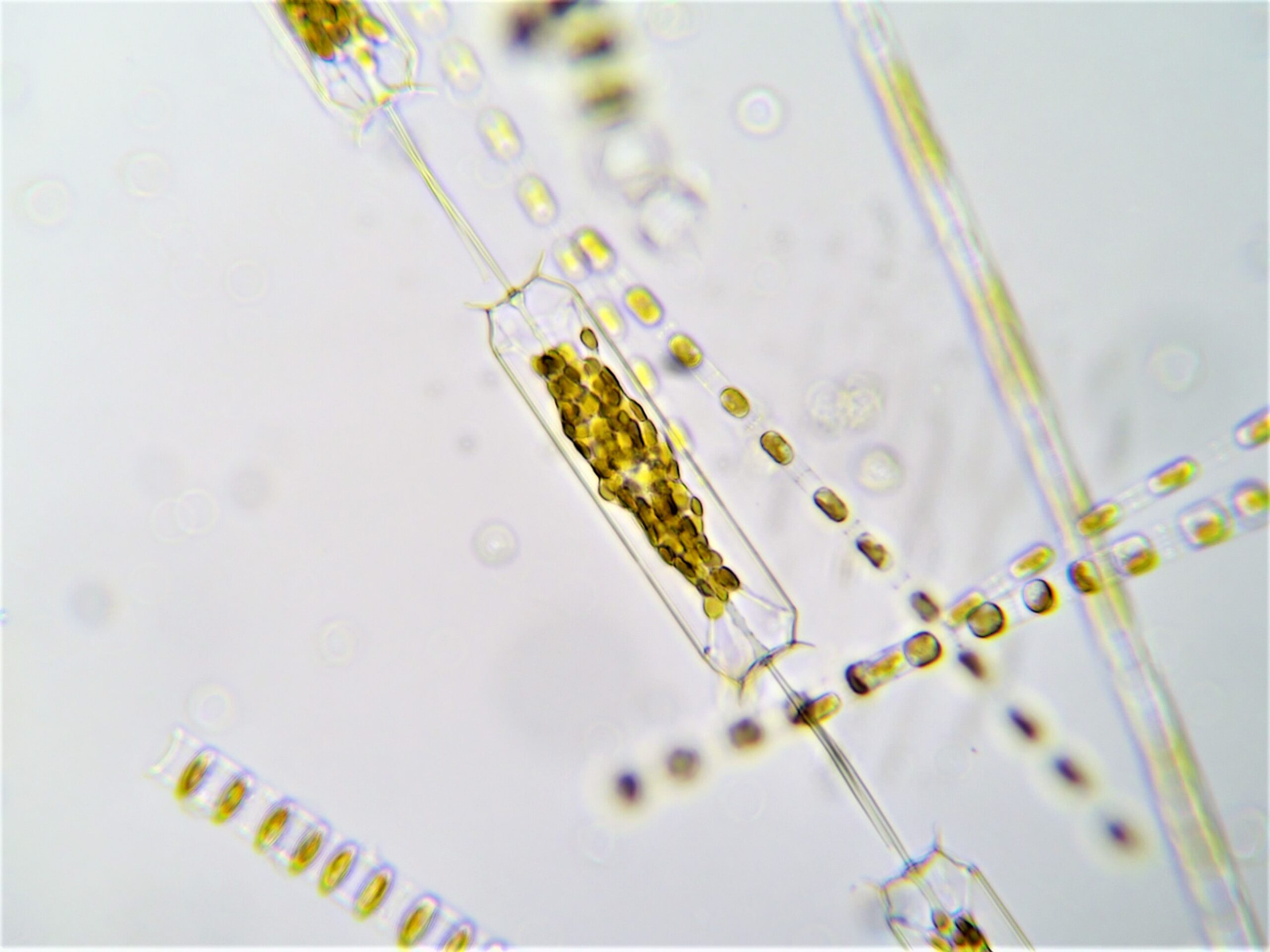
For example, the microscope we used to take these pictures is pretty good and shows a lot of detail, usually enough to make a solid genus level identification, but not all the detail we need to make a species level identification. This means that while we usually know the group that a diatom under the scope belongs to, we might not have enough information to determine its species. In this scenario, we would label the organism as Ditylum spp., aka a diatom in the Ditylum genus that we aren’t sure the exact species of, in the same way that sometimes we might see a bird flying over the water, and we know its a type of seagull, but we don’t know for sure if its a herring gull or a laughing gull.
With spring finally here and the plankton bloom well underway in the Hudson River, our River Project team has been analyzing these samples regularly. If you’ve enjoyed this closer look at how we study building blocks of the Hudson River food chain, stay tuned for more learning opportunities this season. We also offer Plankton Microscopy Field Trips, and you can learn more about those here.